 The heel is one of the places in our body that bears the most weight daily. When the heel is injured, like in a fracture, it can make normal activities like walking difficult. In addition to the pain of a broken heel, the bone itself may widen, shorten, or become otherwise deformed without proper treatment. Because of the severity of these injuries, treatment can often involve surgery and other invasive options, and delays in treatment can mean a worse outlook.
Anatomy of the Heel
Our feet contain many small bones, but they are typically divided into three parts: the hindfoot, the midfoot, and the forefoot. The hindfoot and midfoot include seven bones, the largest of which is the calcaneus or heel hone. It lies at the back of the foot below three other bones that make up the ankle joint, including the talus or a small foot bone that works as a hinge between bones in the legs.
The talus and the calcaneus form the subtalar joint, which allows side-to-side movement of the hindfoot. This joint is critical for balancing on uneven surfaces and other lateral movements while walking.
The calcaneus is often compared to a hardboiled egg because it has a thin, hard shell on the outside with a softer, spongy bone on the inside. If the outer shell is broken, the bone usually collapses and becomes fragmented.
There are various forms of calcaneal fractures. When the joints are involved, a fracture can involve damage to the cartilage- this is the most severe form of the injury. When fractures do not involve the joint, it is more likely to be a crushed bone or a small piece of bone pulled off of the calcaneus.
Causes of Heel Fractures
In most cases of a broken heel, the calcaneus is fractured as a result of a fall from a height where the person lands on their heel or another high-impact trauma like a car accident. The type and severity of fractures can vary, but this injury usually involves some impact.
Fracture patterns are similar in different cases. Some mechanisms that can lead to a fracture include:
If you fall and land on your feet, the weight of your body is directed downward and placed onto your heel. This can drive the talus bone directly into the calcaneus, causing the injury.
In a car crash, the calcaneus can be driven up against the talus when the heel is crushed against the floorboard.
As a general rule, the more impact, the more damage caused. In some of these high-energy impacts, you may see associated injuries like spine, hip, or heel fractures.
In rare cases, stress fractures can occur in the heel due to overuse or repetitive stress on the bone.
Signs of a Fractured Heel
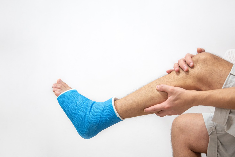
The primary sign of a broken heel is pain, especially pain that worsens during walking or otherwise placing pressure on the heel. With some minor fractures, the pain may cause you to limp but not be enough to prevent you from walking as the Achilles tendon supports your body weight. Pain may be accompanied by bruising and swelling of the foot.
More severe injuries can result in deformity of the heel. In these cases, you may see changes to the shape of your foot externally. This can also cause you to struggle with walking, as the muscle and tendon can’t generate enough energy to support your weight. You may feel like your foot and ankle are unstable, causing you to walk improperly or trip often.
Diagnosing a Broken Heel
 If you are presenting with the symptoms of a broken heel, your doctor will first discuss the circumstances of any injuries with you. For example, if you fell from something like a ladder, they may want to know how far you fell, what position you landed in, and what material you landed on. The more details you provide, the more accurate your diagnosis will be. They may also ask about other medical problems, your medications, lifestyle habits like smoking, and injuries you have suffered in the past.
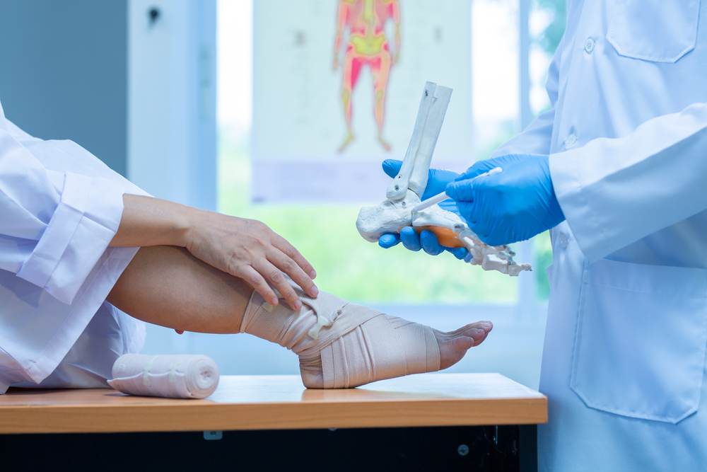
The first thing an orthopedic doctor will do is perform a physical examination of your heel. This may involve examining your foot and ankle to find skin that was damaged or punctured, as well as taking the pulse of your foot in various spots to ensure a good blood supply to the toes. You may be asked to move your toes and whether you can feel certain sensations on the bottom of your feet. If you have any other potential injuries, the physical examination can look at the rest of your leg, pelvis, and spine.
If a calcaneus fracture is suspected after a physical examination, imaging studies can confirm the diagnosis and determine the severity of the fracture. The most common form of scanning will be an x-ray, which creates an image of the bone, including any breaks or displacement.
Because of the complex anatomy of the calcaneus, it is possible that a CT scan may be needed after an x-ray has confirmed the break. This scan can produce a more detailed image of your foot, which will give your doctor more information on the severity of your fracture.
Treatment for Heel Fractures
 When determining the best way to treat a broken heel, your doctor will consider a number of factors such as the cause of your injury, the severity of the break, your overall health, and whether you suffered additional soft tissue damage. Because of the frequency with which bones widen after fracture, the goal of treatment is usually to restore the heel’s normal anatomy. If this is possible, you will work toward a better outcome and the ability to use your heel as normal.
Non-Surgical Treatment
If the force of the injury has not displaced the pieces of your broken bone, your doctor can explore non-surgical options. The most common is immobilization, using a cast, splint, or brace to hold your foot in position while it heals. You might have to wear a cast for 6 to 8 weeks or longer, during which time you will not be able to put any weight on your foot.
Before you can be put into a cast or brace, you may need to use the RICE technique. This technique has four components:
- Rest: Stay off the injured foot as much as possible.
- Ice: Use cold temperatures to reduce swelling and pain. A bag of ice or ice pack wrapped in a thin towel can be applied to the heel in 10-20 minute spurts.
- Compression: Wrap the foot in an elastic bandage or use compression socks.
- Elevate: Keep the foot at or above the level of your heart.
This can help manage pain and prevent further injury before you can be treated.
Surgical Treatment
It is very common for broken heels to require surgery to restore the normal anatomy of the foot. If the bone has shifted out of place or become displaced, surgery will be recommended to restore this normal shape. This decision is usually about managing risks. While surgery comes with risks of infection, nerve damage, or problems healing, an untreated fracture can lead to long-term issues like pain, arthritis, or a limp.
If the skin around your fracture hasn’t been broken or punctured, you may be told to wait for surgery until swelling has gone down. Elevating your leg and keeping it immobilized will help this process happen, which also gives injured skin the chance to recover. The ability to wait for surgery can lead to a better overall recovery and a lower risk of infection.
However, open fractures can expose the injury to the environment and have a high risk of infection. These injuries will need to be treated immediately with surgery to clean the wound and remove the damaged tissue. Early surgery can also be necessary if a piece of the calcaneus is pulled off in an avulsion, when the Achilles tendon splits away from the bone. Emergent surgery in these cases can risk injury to the skin around the Achilles tendon.
Different surgical procedures will be necessary for different types of fractures. The two most common are:
- Percutaneous screw fixation: When bone pieces are large, they can move back into place without making a large incision. Doctors can insert special screws through small incisions to hold the fracture together and allow the bone to heal.
- Open reduction and internal fixation: An open incision can be made to reposition the bones into their normal alignment. Wires, metal plates, or screws can be used to hold the bone in place.
Rehabilitating a Broken Heel
Whether your treatment is surgical or non-surgical, some form of rehabilitation is needed before you can return to daily activities. Depending on the severity of your fracture and other injuries, you may not fully return, but the goal will be to restore as much motion and strength as possible.
Some patients will be able to begin placing weight on the heel again after a few weeks, but 3 months is typically recommended as the amount of time to wait. Partial weight-bearing may begin earlier. In the early recovery period, your doctor may encourage foot and ankle motion when it does not cause too much pain.
Physical therapy is an important part of this rehabilitation. A therapist may provide specific exercises to improve your range of motion and strengthen the muscles that support your foot and ankle. These exercises may be painful or difficult at the beginning, but they will become easier over time as you heal.
A physical therapist may also provide assistive devices, like crutches, a cane, or a walker when you begin walking again. Placing too much weight on the foot could cause your bones to move out of place and necessitate another surgery or cause screws that were placed to loosen and break. If you are advised to use any kind of device to prevent too much weight on your heel, be sure to follow your doctor’s directions exactly. Orthotics may be recommended even after you are walking to help prevent further issues.
Outcomes and Potential Complications
For those with minor injuries, like a crack in the bone with little muscle damage, normal activities could resume as quickly as 3 to 4 months after surgery. More severe cases could take 1 to 2 years before recovery is complete. It is not common for patients to regain full range of motion in their foot and ankle after severe injury, though usually enough is restored to continue on with daily life. However, those whose job or leisure activities involve a lot of walking or climbing may experience major changes to their lifestyle or career.
Without proper treatment and follow-up, complications are common with a calcaneus fracture. Minor complications may include delayed wound healing, nerve irritation, tendon irritation, joint stiffness, chronic pain, and chronic swelling. These can often be managed alongside treatment of the injury.
Major complications can result from surgery, therapy, or the injury itself. Failure of a wound to heal and infection are the most common complications, though post-traumatic arthritis is also possible. Additional surgery may be required in some cases.
The best way to avoid these complications is to seek treatment early and follow any guidance given by your doctors. They will work with you to ensure the least risk of problems and identify any signs of a complication early so that treatment can be personalized.
At AICA Marietta, our goal is to help you reach the best outcome possible. Whether you suffered serious trauma to the heel or suspect a stress fracture, our team of specialists will be able to diagnose the condition and provide an individual plan for healing. Contact us today to begin the process of treatment with our experts!